Mechanics of Morphogenesis:
The Biology and Engineering Behind the Mysteries of Embryonic Development.

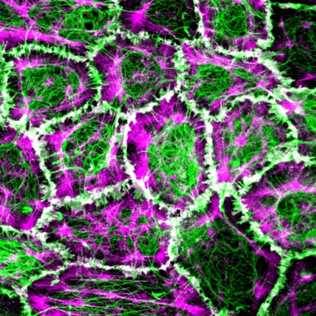
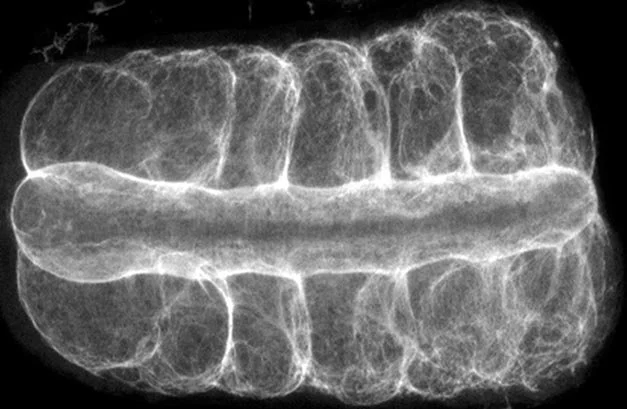
Physics Matters! Biological processes play out in the physical world where work, energy, and kinetics dictate the shape and form of embryos and organs.

Our mission is two-fold, first, to expose the ways in which the environment, genome, cell biology, and mechanics are integrated during development, and second, to turn the principles of development into practical technologies for tissue engineering.

How physical forces shape developing embryos.
How embryos build their organs and body plans remains one of the big open questions in biology. Modern genetics has shown that many animals use very similar genes, proteins, and signaling pathways to control development, yet the exact physical processes that turn these shared genetic programs into very different working structures have been harder to pin down.
This research program helped launch the field of developmental biomechanics and is now revealing how genes link to the forces and mechanical processes that change embryo shape. This work began with Davidson’s PhD studies on sea urchin gastrulation and has continued to grow since then.

How cells and molecules drive large-scale tissue shape change.
Many of the key links between genes and the physical forces that shape tissues show up at the level of single cells. By watching how cells move on their own and in groups during morphogenesis, it becomes possible to see how specific proteins set up mechanical behaviors in the embryo.
Work from the MechMorpho group has revealed how signaling systems, such as planar cell polarity pathways, are tied to force-generating cell activities and new kinds of cell movement. This research shows that cell behavior is not just the end-point of simple “gene A activates gene B” chains, but reflects integrated systems that coordinate multiple physical and molecular processes across a tissue.

Computer models of how cells and embryos develop.
Lance left industry to pursue a PhD in biophysics, aiming to build advanced computer models of how cells and embryos develop. As he worked, it became clear that there were too few physical measurements on embryos, so the project grew into a combined effort in theory and experiment, focused on biomechanics and biophysics.
The MechMorpho/Davidson group and its collaborators now develop computer models that link detailed knowledge of the cytoskeleton and cell adhesion with how tissues generate and respond to forces. More recently, the lab has begun to weave these models together with machine learning and systems bioengineering tools.

How the extracellular matrix shapes developing vertebrate embryos.
Extracellular matrix (ECM) has long been a central focus in developmental biology. In early vertebrate and mammalian embryos, ECM proteins such as fibronectin and laminin are found almost everywhere and surround many types of cells.
Genetic studies that remove ECM components or their receptors, or weaken their function, show that ECM is essential both for setting up correct cell identities and for organizing the layered structure of the early embryo. These studies also reveal that ECM helps the embryo “self-assemble” mechanically, guiding how tissues move and fit together.
During Lance’s postdoctoral work in the frog Xenopus laevis, he mapped when and where fibronectin matrix forms in the embryo and tested how fibronectin and its receptors control groups of cells migrating together. Ongoing work in the lab examines how ECM shapes tissue-scale mechanics and coordinated cell movements during the earliest stages of development.

Studying how tissues take shape in embryos, using frog (Xenopus) as a model.
Xenopus laevis frog embryos are a leading model for studying how embryos change shape and form tissues. Its external fertilization, large eggs, rapid development, and easy care make it ideal for learning core ideas in experimental embryology. The large, synchronously developing embryos provide abundant material for biochemistry and modern genomic and bioinformatic studies. Because the embryos can be cut and recombined and still develop well, they are also powerful tools for probing the physical mechanics of vertebrate development. Work in Xenopus continues to reveal basic biological principles and to inspire new translational approaches in medicine and biotechnology.

Building tools that use ideas from embryonic development to grow new tissues.
Arthur C. Clarke wrote that “any sufficiently advanced technology is indistinguishable from magic.” Embryonic development has that same sense of mystery, but it follows rules that can be understood. This work aims to turn those rules of developmental biology into a practical engineering toolkit for building tissues.
The lab partners with other groups to test and refine design principles that tissue engineers can actually use. Using microfluidics and microfabrication, the team can steer complex programs of tissue shape change and self-organization. The long-term goal is to “forward engineer” how tissues assemble themselves, so that the power of developmental biology can be harnessed for real-world therapies.